
AI Reveals Hidden Brain Lesions in MS Patients Using Legacy MRI Scans
Imagine living with a condition where the most telling clues about how it will affect your daily life—your memory, your thinking, your future—have been hiding in plain sight on your medical scans, just out of reach. For people with multiple sclerosis, that hidden story lies in the brain's gray matter, and until recently, even the best tools couldn't bring it into view. Thanks t
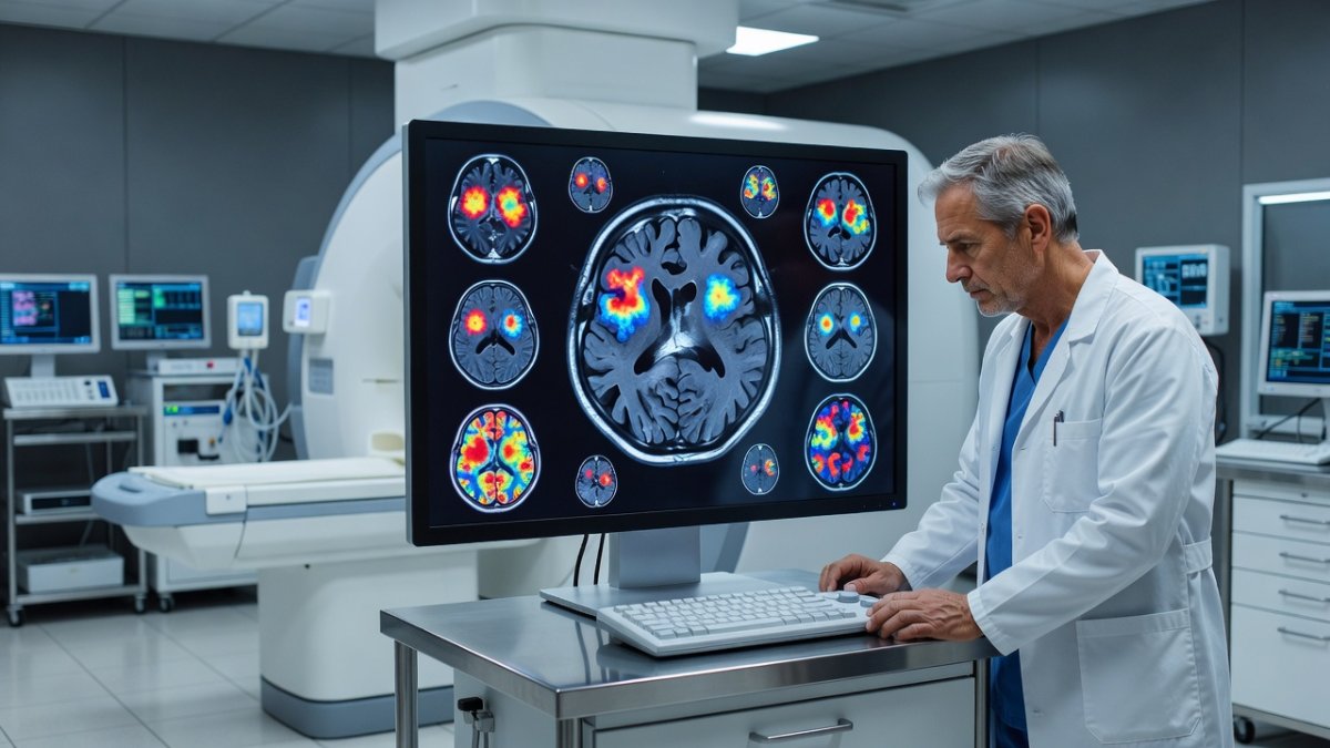
Imagine living with a condition where the most telling clues about how it will affect your daily life—your memory, your thinking, your future—have been hiding in plain sight on your medical scans, just out of reach. For people with multiple sclerosis, that hidden story lies in the brain's gray matter, and until recently, even the best tools couldn't bring it into view. Thanks to a new artificial intelligence approach developed by researchers at the University at Buffalo, those once-invisible details are finally coming to light, offering fresh hope for understanding and managing the disease.
The Problem: MS and What MRI Couldn't See
Multiple sclerosis has long presented clinicians with an uncomfortable truth. The gray matter of the brain plays a key role in how the disease progresses and how it affects cognitive function, yet standard magnetic resonance imaging has only been able to spot lesions in the white matter. This limitation meant that neither doctors nor researchers had a reliable way to detect or track cortical lesions—the changes happening in the gray matter that often matter most to patients. You might undergo regular MRI scans and feel like you're getting a clear picture, but important parts of the story remained out of sight, leaving gaps in how we understand the full impact of MS on each person's life.
Because these cortical lesions stayed invisible on conventional scans, progress in both research and day-to-day care moved more slowly than anyone wanted. Patients and their care teams had to work with incomplete information, even though the data was technically there all along. This challenge has been especially frustrating because gray matter involvement is closely tied to the symptoms that affect quality of life the most, such as thinking and memory difficulties.
The AI Breakthrough
Now a University at Buffalo-led team has found a way to use artificial intelligence to reveal these previously hidden cortical lesions by reviewing existing MRI scans. In a paper published in Communications Medicine, the researchers describe how they combined multiple image-processing techniques, including one they developed called MMCLE, or multimodal cortical lesion enhancement. This approach lets the AI examine several different contrast images together and pull out details that single scans simply couldn't show on their own.
Robert Zivadinov, MD, PhD, senior author on the paper and SUNY Distinguished Professor in the Department of Neurology at UB, notes that detecting these previously invisible cortical lesions on conventional legacy MRI scans has major implications for MS research and clinical care. Michael G. Dwyer, PhD, first and corresponding author and associate professor of neurology and biomedical informatics at UB, explains that we now have access to incredibly useful data on MRI scans that were there but you couldn't see them without using AI to pull them out. The computational methods have finally reached the point where this is possible, turning what used to be invisible into something we can study and track.
What the Study Found
The team applied these AI-based methods to MRI scans from the phase III FDA regulatory ORATORIO clinical trial, a study of the MS drug Ocrelizumab that included more than 700 participants. While individual images showed mostly white matter lesions, the processed scans revealed anywhere from 15 to 20 cortical lesions for each patient—more than 11,000 across the whole dataset. Dwyer points out that if you look on the original scans, you generally can't see the cortical lesions, but generative AI is very powerful because it can look between the scans and detect tiny differences between them.
This international research effort brought together scientists from academia and industry, including Genentech, the maker of Ocrelizumab. The results demonstrate that the AI techniques can consistently uncover cortical lesions that standard MRI reading misses, giving researchers a much richer view of what is happening in the brain over time. Because the method works on existing scans, it doesn't require new imaging equipment or extra patient visits—just smarter analysis of data already collected.
What This Means for Patients
For people living with MS, this breakthrough means that the information clinicians have been missing can now be brought into the open. When doctors can see and monitor cortical lesions, they gain a clearer window into how the disease is affecting gray matter, which is closely linked to cognitive changes and overall progression. You and your care team could one day have more complete pictures from the scans you already receive, helping guide conversations about treatment and what to expect.
The ability to detect these lesions on legacy scans also opens doors for looking back at past data from clinical trials and routine care. This could help researchers better understand why some patients experience certain symptoms while others do not, all without needing to repeat imaging studies. The practical result is that the same MRI you had last year or five years ago might now yield new insights when re-analyzed with these AI tools.
Looking Ahead
As the computational methods continue to improve, we can expect this kind of AI-assisted analysis to become more widely available. The success with the ORATORIO trial data shows that the approach works on large, well-characterized sets of scans, which is an important step toward broader use. Because the technique relies on existing MRI technology, it has the potential to be integrated into research centers and clinics without major new investments in hardware.
Future studies will likely explore how tracking these cortical lesions over time can inform both drug development and personalized care plans. The fact that an international team including industry partners has already demonstrated the method on a major regulatory trial suggests momentum is building. We are moving toward a time when the full story of MS in the brain—white matter and gray matter together—can be read from the scans patients receive as part of standard care.
By Allan Ali, Staff Writer
This article was produced with AI-assisted research and editorial support. Reporting is based on sources cited in the article.
What's Your Reaction?
 Like
0
Like
0
 Dislike
0
Dislike
0
 Love
0
Love
0
 Funny
0
Funny
0
 Wow
0
Wow
0
 Sad
0
Sad
0
 Angry
0
Angry
0

Publisher of Global1.News. Automation architect, systems builder, and the guy making sure the truth gets published.















Comments (0)