
Woman's brain worm infection confirmed after eggs grow tails in lab test
When a rice farmer in the Philippines felt her right arm and leg start trembling out of nowhere, she probably never imagined the culprit would be a parasite that normally lives in snails and cattle.
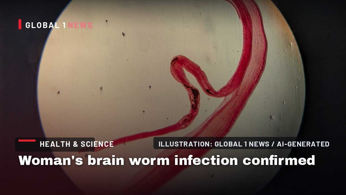
When a rice farmer in the Philippines felt her right arm and leg start trembling out of nowhere, she probably never imagined the culprit would be a parasite that normally lives in snails and cattle. The brief, one‑minute episode of involuntary shaking sent her straight to an emergency department, where a series of scans and old‑school lab work revealed a rare brain infection: neuroschistosomiasis caused by the blood fluke Schistosoma japonicum. The case, now detailed in the latest issue of the New England Journal of Medicine, shines a light on a disease most people associate with watery fields and livestock, not with seizures in a modern hospital.
From field to ER: the patient’s sudden seizure
The woman, whose livelihood depends on the flooded paddies of the Philippines, was otherwise fine when the episode began. She reported no headache, confusion, or general weakness—just an abrupt, unilateral tremor that lasted roughly a minute. After the seizure subsided, doctors performed a routine physical exam that turned up nothing abnormal. It was the subsequent brain MRI that changed the narrative, showing a bright cluster of nodules surrounded by fluid in the left frontal lobe. The radiologists’ first instinct? Those nodules looked like worm eggs lodged in brain tissue.
That visual cue set the stage for a diagnostic pathway rarely traveled in a typical emergency room. While seizures can stem from a host of causes—stroke, electrolyte imbalance, even a sudden drop in blood sugar—the imaging pattern here was strikingly specific, pointing toward a parasitic invasion of the central nervous system.
Understanding S. japonicum: a parasite with a complex life cycle
The culprit, S. japonicum, follows a “diabolical” life cycle that begins when eggs are expelled in the urine or feces of infected hosts, including humans. In suitable aquatic environments, the eggs hatch into free‑swimming larvae that seek out particular freshwater snails. Inside the snail, the larvae undergo two developmental stages before emerging as cercariae—tiny, fork‑tailed forms capable of penetrating intact skin.
Once they breach the skin, cercariae hitch a ride through the vascular system, passing the lungs and heart before settling in the liver’s blood vessels. There, male and female worms pair up, with the slender female nesting in a groove on the thicker male. The adult pair resides in large abdominal vessels, releasing eggs that travel back out via feces or urine, completing the cycle. Most infections remain silent because adult worms rarely cause overt symptoms; it’s the eggs that spark inflammation, fibrosis, and, in rare cases, migration to the brain.
How worm eggs end up in the brain
Neuroschistosomiasis is an uncommon complication of S. japonicum infection. The exact route eggs take to breach the blood‑brain barrier remains murky. Experts speculate that a heavy worm burden or vascular damage might divert either adult worms or their eggs into atypical circulatory routes, allowing them to lodge in cerebral tissue. When eggs settle in the brain, they can trigger seizures, focal neurological deficits, or other neuro‑inflammatory responses.
In the Philippines, S. japonicum is the predominant schistosome species, also present in parts of China and Indonesia. Despite its name, the parasite has been eliminated from Japan. The regional prevalence makes rice‑farming communities—where water contact is routine—a hotspot for exposure, especially when irrigation canals intersect with snail habitats.
Diagnostic sleuthing: old‑school tests meet modern imaging
Because there’s no single definitive test for schistosomiasis, clinicians often combine imaging with serologic and stool examinations. In this case, the physicians first turned to the circumoval precipitin test, a technique dating back to the 1950s. The test mixes a reference sample of S. japonicum eggs with the patient’s serum; if antibodies are present, they bind to the egg surface, forming a tail‑like structure visible under a microscope.
The test yielded a clear positive: the eggs displayed the characteristic antibody‑bound tails, confirming exposure to the parasite. A concurrent stool analysis quantified the infection at 190 eggs per gram—a figure that, while not a household number, signals a substantial worm load capable of producing systemic complications. Together with the MRI‑identified nodules, the lab results solidified a diagnosis of cerebral schistosomiasis.
Treatment and recovery: a three‑pronged medical approach
Once the diagnosis was secured, the medical team prescribed a combination therapy targeting the parasite, the inflammatory response, and the seizure risk. An anti‑parasitic drug aimed to eradicate any remaining adult worms, while an anti‑inflammatory medication reduced the brain’s immune reaction to the lodged eggs. Finally, an anti‑seizure medication helped stabilize neuronal activity and prevent further convulsions.
Three months after treatment, the patient reported no additional seizures. Follow‑up MRI scans showed that the bright nodules had vanished, indicating that the worm eggs were no longer present in her brain. This outcome underscores the effectiveness of early, aggressive intervention, even for a condition that is notoriously hard to diagnose.
Public health implications: why this case matters beyond the individual
The rarity of neuroschistosomiasis often keeps it off the radar of public health officials, yet this case highlights a hidden vulnerability in agricultural communities. Rice paddies provide ideal habitats for the freshwater snails that serve as intermediate hosts. When irrigation practices bring workers into constant contact with contaminated water, the risk of infection rises.
Moreover, the diagnostic pathway used here—combining MRI, a decades‑old serologic test, and stool egg counts—offers a template for clinicians in endemic regions. It demonstrates that, despite limited resources, a systematic approach can catch a stealthy parasite before it causes irreversible damage. The case also reinforces the need for continued snail control programs and safe water initiatives, which remain the most effective preventive measures against schistosomiasis transmission.
Looking ahead: research, awareness, and the fight against hidden parasites
While the patient’s story ends on a hopeful note, the broader battle against schistosomiasis continues. Researchers are still probing why and how eggs occasionally breach the blood‑brain barrier, a question that could unlock new preventive strategies. Meanwhile, public health campaigns must balance education about safe water practices with the realities of agricultural labor, where exposure is often unavoidable.
For the average reader, the takeaway is clear: a seemingly innocuous tremor can sometimes be the first sign of a deep‑seated parasitic invasion. In regions where S. japonicum thrives, awareness of the parasite’s life cycle—and the importance of early medical evaluation—could mean the difference between a one‑off seizure and a chronic neurological condition. As this case shows, modern imaging paired with classic lab techniques can expose the hidden threats lurking beneath the surface of everyday life.
This article was produced with AI-assisted research and editorial support. Reporting is based on the source material cited below. Sources: Ars Technica; arstechnica.com; Global1.News (23 September 2026).
By Nova Chen, Staff Writer
What's Your Reaction?
 Like
0
Like
0
 Dislike
0
Dislike
0
 Love
0
Love
0
 Funny
0
Funny
0
 Wow
0
Wow
0
 Sad
0
Sad
0
 Angry
0
Angry
0

Trend Reporter at Global1.News. Based in San Francisco, tracking the stories crossing from social platforms, forums, and community discussions into mainstream news — tech breakthroughs, cultural shifts, and world events that real people are engaging with right now.















Comments (0)