
Doctors pull knife from man's eye that went through his brain—and he's fine now
When a man in his twenties staggered into a UK emergency department with the hilt of a knife still protruding from his face, the scene could have been ripped from a horror film.
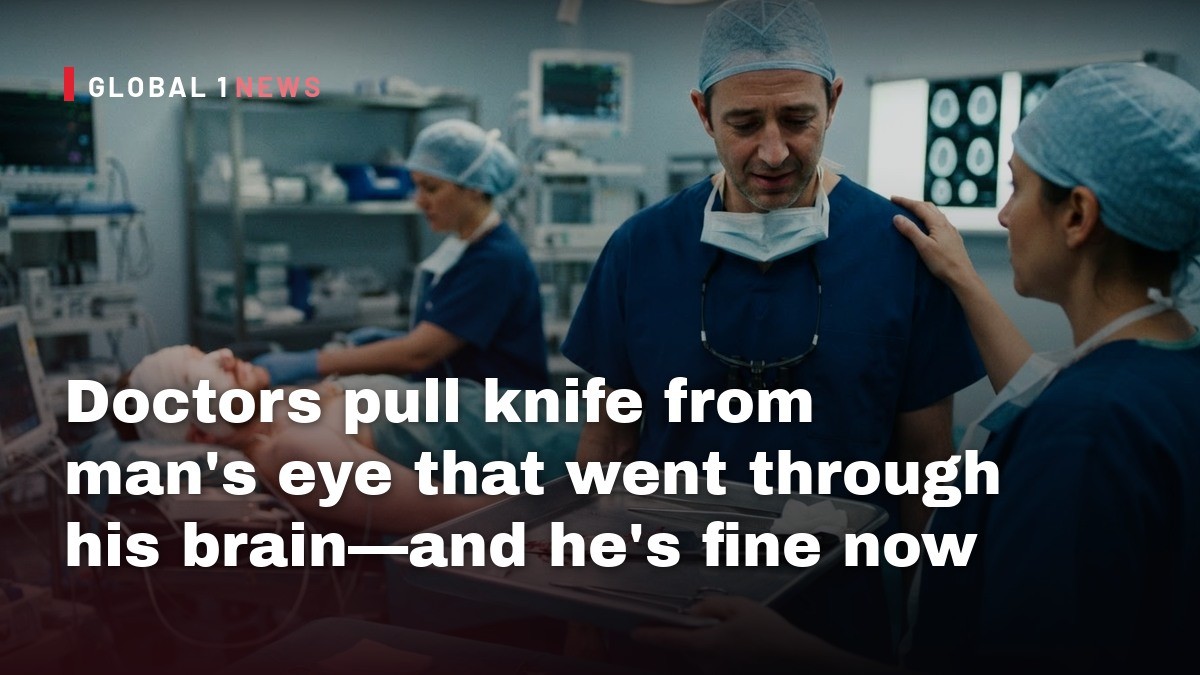
When a man in his twenties staggered into a UK emergency department with the hilt of a knife still protruding from his face, the scene could have been ripped from a horror film. Yet the reality, as documented in a recent BMJ Case Report, was a stark reminder that modern trauma care can turn the unimaginable into a survivable ordeal. The 15‑mm‑wide blade had sliced clean through his right eye, pierced the left frontal and temporal lobes, and lodged near the top of his skull—all while the patient remained conscious, able to speak, and move all four limbs. This case, while extraordinary, offers a valuable lens on how low‑velocity penetrating brain injuries are managed, why outcomes can vary dramatically, and what the medical community can learn from a brush with death.
From the Moment of Arrival: Initial Assessment and Stabilization
Upon arrival, the patient’s condition was paradoxically stable despite the gruesome trajectory of the weapon. He was alert, could answer questions, and demonstrated purposeful movement, though he exhibited agitation—a common response in traumatic brain injury. Clinicians quantified his neurological status using the Glasgow Coma Scale, assigning a score of 12 (E3 V4 M5). This placed him in the “moderate” injury category, just a point above “mild,” indicating that while his brain function was compromised, critical brainstem reflexes remained intact.
Given his confused and agitated state, the medical team faced an immediate dilemma: the risk that he might attempt to extract the knife himself, potentially causing catastrophic hemorrhage. To preempt this, they swiftly sedated and intubated him, securing his airway and preventing further self‑inflicted trauma. Prophylactic antibiotics, including tetanus coverage, were administered to guard against infection—a standard precaution in penetrating injuries where foreign material traverses sterile tissue.
Imaging the Intracranial Path: Mapping the Blade’s Journey
High‑resolution imaging was the next critical step. CT scans revealed that the blade had traversed the right orbit, pierced the left frontal and temporal lobes, and rested against the inner table of the skull. Remarkably, the weapon missed the corpus callosum—a dense bundle of fibers linking the brain’s hemispheres—as well as the left internal capsule, another crucial conduit for motor and sensory pathways. Avoiding these structures likely spared the patient from more severe deficits such as paralysis or profound language impairment.
Vascular assessment showed the blade skirting the left internal carotid artery without breaching it, yet a branch of the middle cerebral artery appeared indented. This close proximity to major blood vessels underscored the precariousness of the injury; a single misstep could have precipitated massive intracerebral hemorrhage, a leading cause of mortality in penetrating brain trauma.
Surgical Strategy: A Conservative “Pull‑and‑See” Approach
After multidisciplinary consultation, neurosurgeons elected a conservative extraction technique. Rather than performing an extensive craniotomy to dissect the blade out, they performed a limited skull opening to visualize the entry point and then employed a “pull‑and‑see” method—essentially sliding the weapon out along its original path. This minimized additional disruption to surrounding brain tissue and reduced the risk of inducing new vascular injury.
Post‑extraction inspection revealed no active bleeding, a testament to the blade’s fortunate trajectory. The wound cavity was irrigated, drained, and the skull defect patched. Unfortunately, the right eye could not be salvaged; the extent of ocular destruction necessitated enucleation. Nonetheless, the preservation of language function and gross motor abilities highlighted how trajectory, more than sheer kinetic energy, dictates outcome in low‑velocity penetrations.
Recovery in the ICU: Rapid Progress and Residual Deficits
The patient’s postoperative course was notable for its speed. Within 24 hours, he was weaned from mechanical ventilation, and his breathing tube was removed. Minor right‑side weakness emerged—a common sign of transient cortical irritation—but resolved within two days, suggesting that the affected motor pathways were not permanently compromised.
Serial imaging tracked a gradual decline in intracerebral air, small hemorrhagic foci, and edema. Importantly, there were no signs of infection, seizures, or cerebrospinal fluid leaks—complications that frequently plague penetrating head injuries. By the time of discharge, the patient was deemed “neurologically well,” though he continued to receive ophthalmologic and mental‑health follow‑up, reflecting the ongoing need to address both physical and psychological sequelae.
Understanding Low‑Velocity Penetrating Brain Injuries
Penetrating brain trauma is typically associated with high‑velocity mechanisms such as gunshots, where kinetic energy creates cavitation and extensive tissue destruction. In contrast, low‑velocity objects—knives, rods, nails—produce a more linear wound track. While the overall mortality is lower, the morbidity can be severe because even a narrow path can intersect vital structures.
The BMJ case report emphasizes that outcomes hinge on the precise trajectory relative to critical vascular and neural substrates. In this instance, the blade’s path spared the corpus callosum, internal capsule, and major arteries, thereby limiting permanent deficits. Nonetheless, the presence of an indented middle cerebral artery branch illustrates that even low‑velocity penetrations carry a non‑trivial risk of vascular injury, underscoring the need for meticulous imaging and surgical planning.
Historical Context: From Phineas Gage to Modern Trauma Care
The case evokes the infamous 19th‑century story of Phineas Gage, whose iron rod traversed his skull and altered his personality. While Gage survived, his post‑injury behavioral changes have become a cautionary tale about frontal‑lobe trauma. Modern parallels, such as a nail‑gun injury cited by the authors, demonstrate that survivability has improved dramatically thanks to advances in imaging, critical care, and surgical techniques.
What unites these cases is the principle that the brain’s functional outcome often depends less on the external severity of the wound and more on which structures are intersected. The recent UK case reinforces this lesson: a knife that could have sliced through the internal carotid artery or the internal capsule would likely have proved fatal, yet its fortunate trajectory allowed for a near‑complete neurological recovery.
Implications for Clinical Practice and Public Awareness
For trauma teams, this case underscores the importance of rapid airway protection, early sedation to prevent self‑extraction, and comprehensive imaging before any operative maneuver. The decision to employ a conservative extraction technique, rather than an aggressive craniotomy, reflects a nuanced balance between exposure and iatrogenic injury—a balance that can be guided by detailed CT angiography.
From a public‑health perspective, the incident also highlights the intersection of mental health and trauma. The patient’s history of depression, noted by his physicians, raises questions about the circumstances leading to such a self‑inflicted or accidental injury. While the exact mechanism remains unclear, the report serves as a reminder that individuals with mental‑health challenges may be at heightened risk for severe self‑harm, and that integrated psychiatric support is essential in both prevention and post‑injury rehabilitation.
In sum, the survival of a young man who walked into a hospital with a knife through his eye and brain is a testament to modern medicine’s capacity to turn near‑fatal wounds into recoverable injuries—provided that clinicians act swiftly, image meticulously, and respect the delicate architecture of the human brain.
This article was produced with AI-assisted research and editorial support. Reporting is based on the source material cited below. Sources: Ars Technica; arstechnica.com; Global1.News (10 November 2026).
By Jessica Ali, Staff Writer
What's Your Reaction?
 Like
0
 Dislike
0
 Love
0
 Funny
0
 Wow
0
 Sad
0
 Angry
0

Editor-in-Chief at Global1.News. Atlanta-based journalist who cuts through the BS and tells it like it is. Lead anchor, host, and the voice you hear when the spin stops and the truth starts.















Comments (0)